
CONE BEAM CT
What is this ?
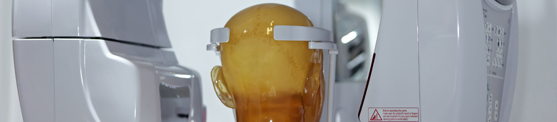
Cone Beam Computed Tomography (CBCT) consists of a rotating gantry along an axis with an x-ray source and detector on opposite sides for acquiring images. The unit rotates in a complete or partial arc along a central fulcrum where the patient6 is positioned. Unlike the conventional Ct which acquire multiple image slices with a fan-shaped beam of ionizing radiation and compiles them together, resulting in a 3D image, CBCT involves a single rotation of the gantry, for the entire image construction.
Why I need this :
Dental imaging involving the CBCT has been proved to be a boon among doctors worldwide, helping in diagnosing acute cases of dental implant, abnormal teeth visualization, evaluation of the jaws and face, cleft palate assessment, diagnosis of dental caries (cavities), endodontic (root canal) diagnosis, and diagnosis of dental trauma.
CBCT acts as a helpful tool in interventional radiology.
Since it provides for 3D images instead of 2D, it is an essential tool in maxillofacial imaging, mandibular canal tracking and imaging of impacted teeth.
Services offered :
- The radiation dose is extremely less, so much so that it does not qualify as a potential health threat.
- The more comfortable standing position of the patient is offered.
- Patients should not have claustrophobia.
- High-resolution images are obtained.
What shall I expect :
- It is advisable to bring a record of your medical imaging history.
- No prior preparation such as fasting or ion tracing is needed.
- This process is completely safe, non-invasive and gleefully painless.